
Congenital Foot Deformities Care
Specialized pediatric podiatry for children born with foot differences, including accessory navicular syndrome.
What You Need To Know
Understanding accessory navicular syndrome and other congenital foot conditions in children.
Accessory Navicular Syndrome
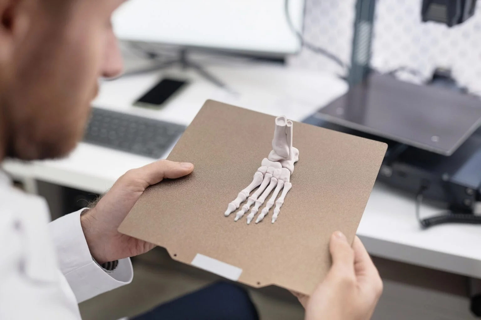
The accessory navicular (os navicularum or os tibiale externum) is an extra bone or cartilage located on the inner side of the foot just above the arch. It is incorporated within the posterior tibial tendon, which attaches in this area and can lead to what’s called Accessory Navicular Syndrome. An accessory navicular is congenital (present at birth) and is not part of normal bone structure, so it is not present in most people.
People who have an accessory navicular are often unaware of the condition if it causes no problems. However, some develop a painful condition known as accessory navicular syndrome when the bone or posterior tibial tendon are aggravated. This can result from trauma such as a foot or ankle sprain, chronic irritation from footwear rubbing against the extra bone, or excessive activity and overuse.
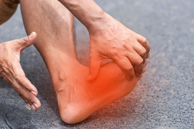
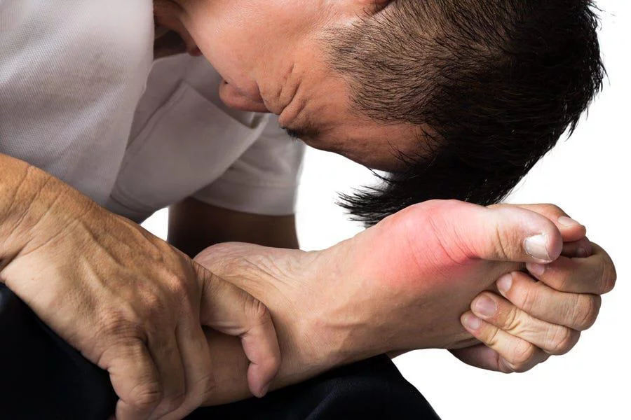
Adolescence is a common time for symptoms to first appear, when bones are maturing and cartilage is developing into bone. Sometimes symptoms do not occur until adulthood. Signs include a visible bony prominence on the midfoot, redness and swelling of that prominence, and vague pain or throbbing in the midfoot and arch during or after activity.
Many people with accessory navicular syndrome also have flat feet, which puts more strain on the posterior tibial tendon and can irritate the accessory navicular. If you or your child are experiencing these symptoms, contact our office to schedule an evaluation with one of our podiatrists.
Diagnosis and Treatment Options
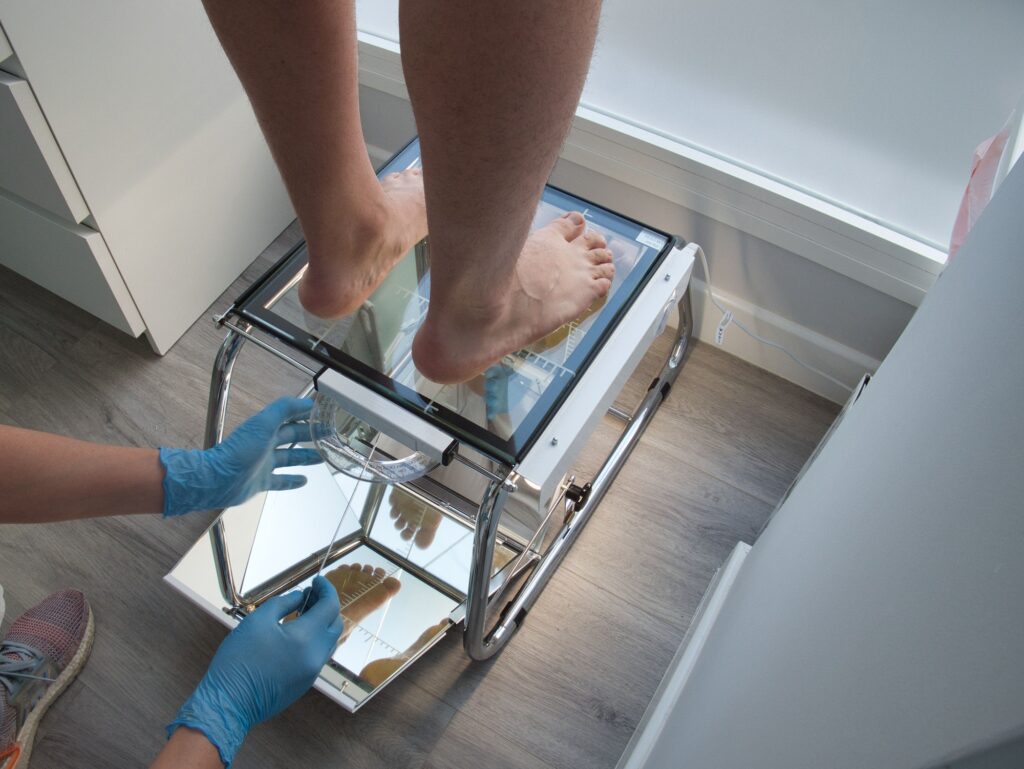
To diagnose accessory navicular syndrome, your doctor will ask about symptoms and examine the foot, looking for skin irritation or swelling. The doctor may press on the bony prominence to assess discomfort. Foot structure, muscle strength, joint motion, and the way the patient walks may also be evaluated. X-rays are usually ordered to confirm the diagnosis, and an MRI or other advanced imaging may be used if pain or inflammation persists.
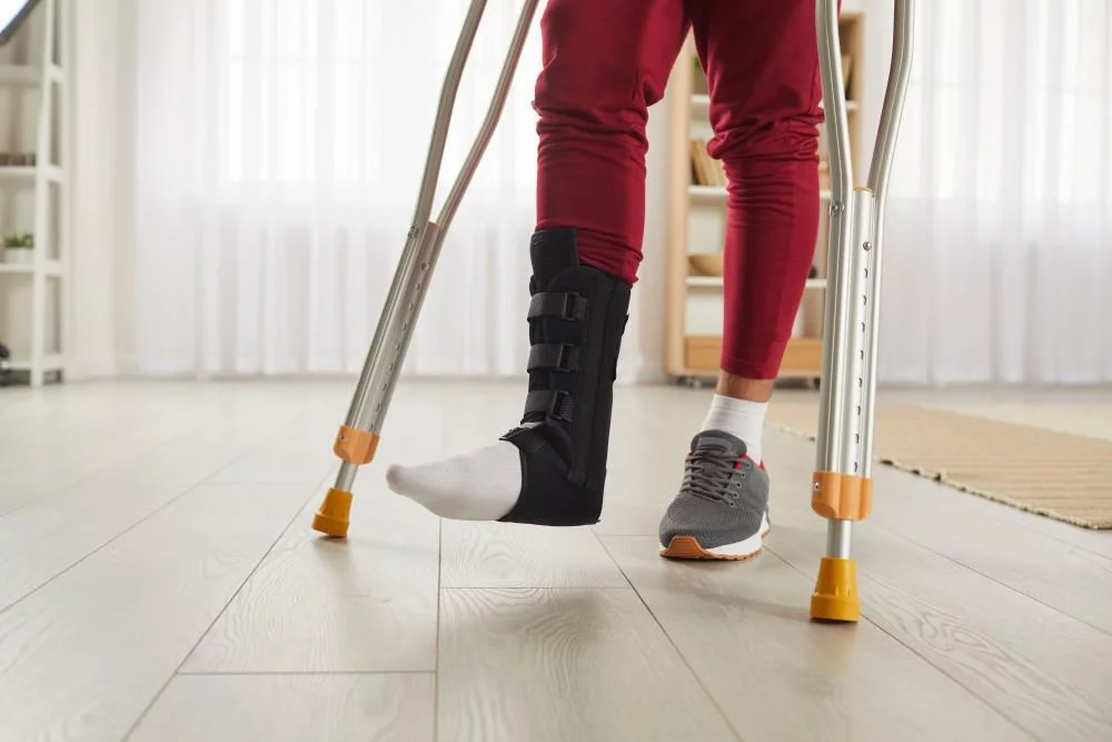
The goal of nonsurgical treatment is to relieve symptoms. Immobilization in a cast or removable walking boot allows the area to rest and decreases inflammation. Ice applied with a thin towel between the bag and skin helps reduce swelling. Oral NSAIDs such as ibuprofen may be prescribed, and in some cases oral or injected steroid medications may be used along with immobilization.
Physical therapy may be prescribed, including exercises to strengthen the muscles and decrease inflammation. These exercises may also help prevent recurrence. Over-the-counter or custom orthotic devices that fit into the shoe provide arch support and may help prevent future symptoms.
If nonsurgical treatment fails, surgery may be appropriate. It may involve removing the accessory bone, reshaping the area, and repairing the posterior tibial tendon. This extra bone is not needed for normal foot function. Our podiatrists will work with you to determine the right treatment path for your situation.
Locations Offering Pediatric Foot Care
Find a Bay Area Foot Care location near you for pediatric podiatry services.
















Concerned about your child’s foot development? Let’s take a closer look.
Our podiatrists are here to help. Schedule a visit at a location near you.